Bean Cell Structure Microscopy Analysis
The Intricate Architecture of Bean Cells: An Overview
Bean cells, like all plant cells, are marvels of biological organization. Their structure is defined by rigid cell walls, chloroplasts for photosynthesis, and vacuoles that maintain turgor pressure. Microscopy unlocks this hidden world, revealing details critical for understanding plant physiology. Unlike animal cells, bean cells feature specialized components such as plasmodesmata—microscopic channels that facilitate communication between cells—and amyloplasts for starch storage. This section explores the foundational elements of bean cell biology, setting the stage for deeper analysis through microscopy.
Preparing Bean Samples for Microscopic Analysis
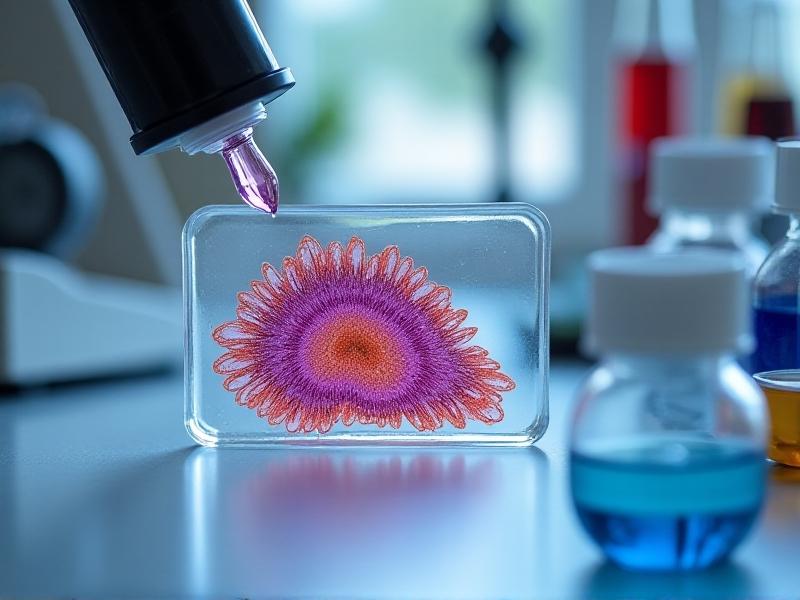
Sample preparation is pivotal for clear microscopy results. Fresh bean slices are thinly sectioned using a microtome to ensure light penetrates the tissue. Staining with iodine highlights starch granules, while toluidine blue enhances visibility of cell walls and nuclei. Fixation with ethanol or formaldehyde preserves cellular integrity. Challenges include avoiding air bubbles during slide mounting and ensuring stains don’t obscure delicate structures. Properly prepared samples reveal crisp organelle boundaries and intracellular details, laying the groundwork for precise observation.
Light Microscopy vs. Electron Microscopy: Comparing Techniques
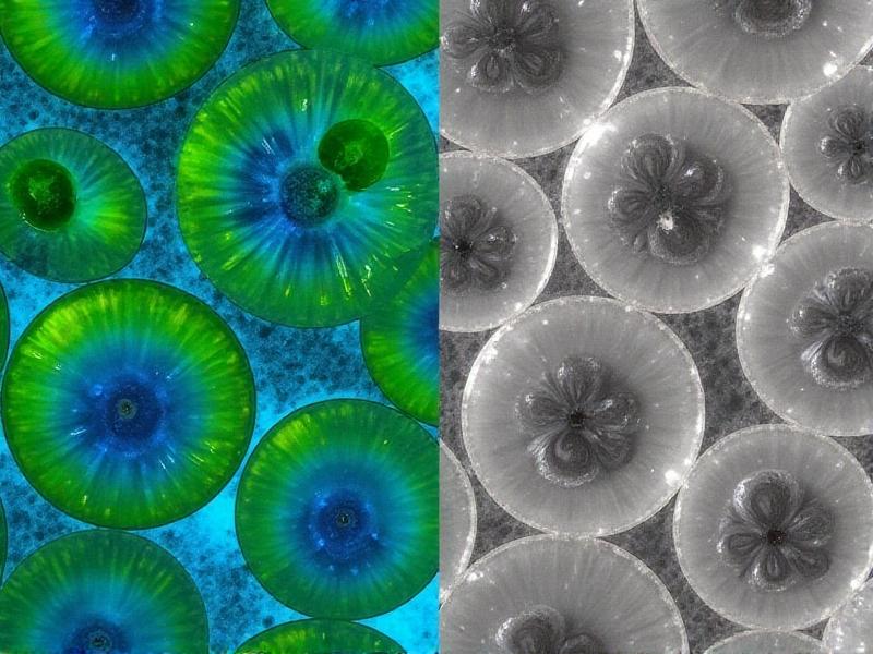
Light microscopy offers live-cell imaging and color visualization but is limited by lower resolution. Phase-contrast variants enhance contrast for organelles like mitochondria. Electron microscopy, while requiring vacuum-sealed samples, provides nanometer-scale resolution, uncovering ultrastructures like thylakoid membranes in chloroplasts. Scanning electron microscopy (SEM) adds 3D surface topography, whereas transmission electron microscopy (TEM) details internal arrangements. Each method has trade-offs: light microscopy is accessible and quick, while electron microscopy demands specialized equipment but delivers unparalleled detail.
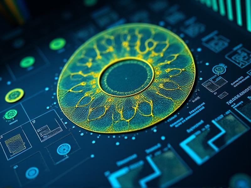
Interpreting Cellular Features Through Image Analysis
Modern software tools quantify cell dimensions, organelle counts, and starch granule distribution. ImageJ or MATLAB algorithms segment cell walls from cytoplasm, measure vacuole volume, and detect abnormalities like plasmolysis. Machine learning models trained on bean cell datasets can auto-identify structures, reducing human error. For example, chloroplast alignment in response to light can be tracked over time. This section discusses how computational analysis transforms raw micrographs into actionable data, bridging observation and insight.
Key Observations in Bean Cell Ultrastructure
High-resolution imaging reveals unexpected complexities. For instance, the endoplasmic reticulum forms dynamic networks around vacuoles, and plasmodesmata exhibit selective permeability. Starch granules vary in density between root and leaf cells, reflecting metabolic demands. Mitochondria cluster near cell walls in active transport zones. These findings challenge simplistic models of plant cells, emphasizing adaptability and functional specialization within bean tissues. Such details inform genetic engineering and agricultural optimization efforts.
Applications in Agriculture and Biotechnology
Microscopy-driven insights into bean cells have real-world impacts. Analyzing cell wall composition aids in developing disease-resistant bean varieties. Studying chloroplast efficiency informs photosynthesis optimization for higher crop yields. Biotech firms use cryo-electron microscopy to observe gene expression in modified beans. Educational kits leverage simple light microscopes to teach students about plant biology. By linking cellular structure to function, microscopy becomes a tool for innovation in food security and sustainable farming.